This article explores whether upright MRI may be a useful tool for evaluating EDS-related spinal instability. Understanding the limitations of standard MRI and what positional imaging could offer instead, may help patients, clinicians, researchers, advocates, and healthcare providers better navigate the complex neurosurgical landscape of EDS.
Why Supine MRI May Miss the Problem
Cervical spine: Unremarkable.
Thoracic spine: Unremarkable.
Lumbar spine: Slight bulging disc at L5-S1.
For many people with Ehlers-Danlos syndrome (EDS), this looks like a typical MRI report: a minor finding that feels disconnected from the severity of their symptoms. Unfortunately, this can be the beginning of years spent searching for answers while symptoms are misunderstood, misattributed, or left unexplained. And while a report like this may leave us feeling dismissed or unheard, the problem isn’t always the clinician, or how they interpret our test results. Sometimes the issue is the test itself.
Standard MRI is one of the most powerful diagnostic tools in modern medicine, and for most conditions, imaging while lying down is entirely appropriate. But when spinal instability is present in EDS, it is often considered a dynamic mechanical problem. A spine may appear relatively normal at rest yet become unstable under the demands of daily life.
When you have a patient population whose symptoms are often aggravated by standing, sitting, and carrying the weight of their own body throughout the day, it seems odd that we rely on a tool designed to capture the spine horizontally at rest. Imaging the spine in its least symptomatic state (lying down) and expecting to identify a positional problem creates a diagnostic mismatch. This reflects a limitation in the way MRI is commonly used, rather than a flaw in the technology itself.
EDS and the Spine
Joint instability can occur in multiple EDS subtypes (such as hypermobile EDS and classical EDS), meaning anywhere along the spine can be affected, from the base of the skull through the neck, mid-back, lower back, and sacrum. And this instability can lead to pain, neurological symptoms, and other complications over time.
What Upright MRI Offers
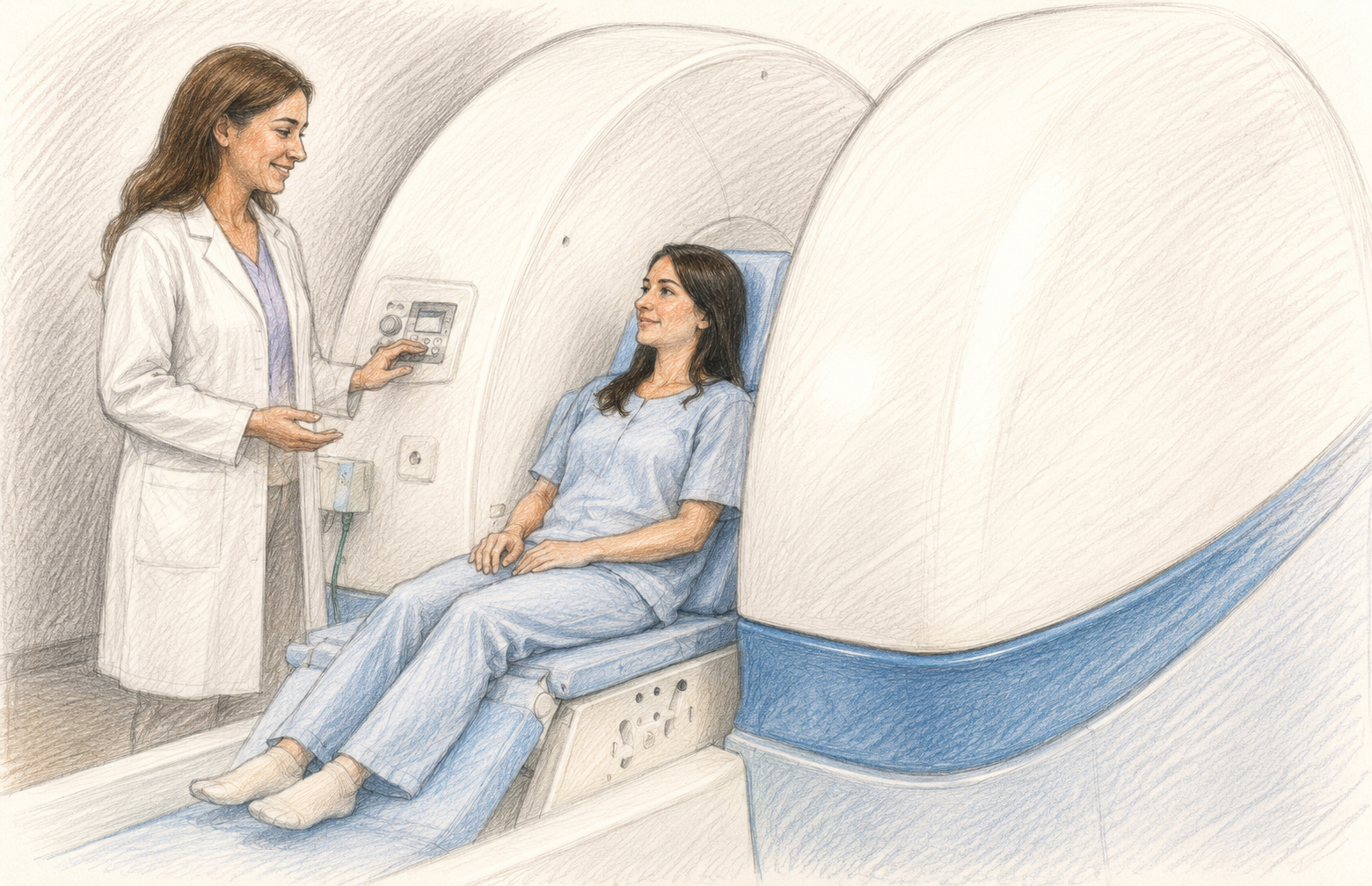
Positional MRI (pMRI), also known as upright or weight-bearing MRI, allows the spine to be imaged while sitting or standing rather than lying down. Some systems can even compare images taken while upright with those taken supine during the same session. Because different positions place different stresses on the spine and supporting ligaments, positional imaging may help reveal instability that is not always visible on a standard MRI.
But there is one trade-off. Upright MRI machines typically operate at lower field strengths (commonly 0.6T to 1.0T) compared to the more common 1.5T and 3T hospital MRIs. This translates into slightly less detailed images than the MRI scanners commonly found in hospitals. However, when evaluating spinal instability, the ability to see how the spine behaves under weight-bearing conditions may outweigh the loss of some image detail.
Why Positional Imaging May Be Worth Considering
A growing body of research suggests that upright, dynamic MRI can reveal posture-dependent changes that can be missed on typical MRI. This may be particularly relevant when symptoms consistently worsen with standing, sitting, bending, head movements, or as the day progresses, yet conventional imaging appears largely unremarkable. For example, a 2025 systematic review identified nine studies demonstrating that dynamic and upright MRI can detect weight-bearing abnormalities. These issues included alterations in spinal alignment, narrowing of the spinal canal, spinal cord compression, cerebrospinal fluid (CSF) flow dynamics, and other abnormalities that become more apparent when the body is upright.
For this reason, positional imaging has attracted interest in conditions where symptoms appear strongly influenced by posture, movement, or mechanical loading of the spine. Research has been particularly active in areas that often affect EDS, such as the lumbar spine, craniocervical instability (CCI), Chiari malformation, and other disorders involving where the base of the skull and upper spine meet, known as the craniocervical junction. As interest in these conditions has grown, some clinicians and organizations, including the CCI Foundation, have started recommending upright MRI as part of the evaluation process.
Research Gaps
However, it is important to recognize that demonstrating posture-dependent changes is not the same as proving improved diagnostic accuracy. While studies have shown measurable differences between upright and supine imaging, large-scale studies directly comparing upright to conventional MRI for EDS-related spinal instability remain limited. As a result, many questions about when upright imaging is most useful, and for which patients, have yet to be fully answered. Therefore, more research is needed.
For now, rather than replacing standard MRI, upright MRI may provide an additional perspective when symptoms and conventional imaging do not seem to match. For some patients, seeing how the spine behaves under weight-bearing conditions may offer information that cannot be captured while lying down.
Access and Practical Considerations
Unfortunately, access to upright MRI is not always straightforward. Upright MRI centers are far less common than conventional MRI facilities, and insurance coverage can vary depending on the indication, provider, and location.
As a result, even when positional imaging seems like a reasonable next step, obtaining it may not be easy. Some patients may need to travel significant distances, pay out of pocket, or explore alternative forms of dynamic imaging recommended by their healthcare team.
For patients interested in positional imaging, it may be worth discussing whether upright MRI is available locally, whether preauthorization is required, and whether other dynamic imaging options may be appropriate.
When Might Upright MRI Be Relevant for You?
An upright MRI might be worth discussing with your healthcare provider if:
- symptoms consistently worsen with standing, sitting, or remaining upright.
- symptoms are triggered by specific head or neck positions.
- symptoms worsen throughout the day as the body bears weight.
- conventional imaging appears largely unremarkable despite significant symptoms.
- symptoms and imaging findings do not seem to match.
Final Thoughts
The spine is not a static structure. It bends, rotates, and bears weight throughout the day. In EDS, these normal demands can expose instability and intensify symptoms.
For a condition in which symptoms are often influenced by position, imaging performed exclusively in the least symptomatic position may not always capture the full picture. Upright MRI is not a replacement for conventional imaging, but it can provide an additional perspective when symptoms and imaging findings do not seem to match.
The technology exists. The challenge now is improving awareness, access, and research so that patients and clinicians have the tools they need to see the full picture.
Key Takeaways
- EDS-related spinal instability is often a dynamic problem that may not be fully visible on standard MRI.
- Conventional MRI may not always rule out mechanical or positional contributors to symptoms.
- Upright MRI allows imaging under weight-bearing conditions and may reveal abnormalities not seen while lying down.
- Positional imaging may be particularly useful when symptoms appear strongly related to posture or movement.
- Research comparing upright MRI to standard MRI remains limited and further research is needed.
- Upright MRI is not available everywhere, and insurance coverage can vary, making access a challenge for some patients.
- If symptoms and imaging findings do not seem to match, discuss with your healthcare provider whether upright or positional MRI may be appropriate for you.
Edited by Jacqueline Teti, Editor-in-Chief
Sources
Health Quality Ontario. (2015). Positional magnetic resonance imaging for people with Ehlers-Danlos syndrome or suspected craniovertebral or cervical spine abnormalities: An evidence-based analysis. Ontario Health Technology Assessment Series, 15(13), 1–24.
Henderson, F. C., Sr., Austin, C., Benzel, E., Bolognese, P., Ellenbogen, R., Francomano, C. A., Ireton, C., Klinge, P., Koby, M., Long, D., Patel, S., Singman, E. L., & Voermans, N. C. (2017). Neurological and spinal manifestations of the Ehlers-Danlos syndromes. American Journal of Medical Genetics Part C: Seminars in Medical Genetics, 175(1), 195–211. https://doi.org/10.1002/ajmg.c.31549
Lohkamp, L. N., Marathe, N., & Fehlings, M. G. (2022). Craniocervical instability in Ehlers-Danlos syndrome—A systematic review of diagnostic and surgical treatment criteria. Global Spine Journal, 12(8), 1862–1871. https://doi.org/10.1177/21925682211068520
Manjila, S., Jayaraj Ranjini, N., Rathore, S., Medani, K., Mani, S., Sideras, P., Kaimal, G., Siravuru, A., & Rayasam, K. (2026). Manjila Chiari Protocol 2.0 (MaChiP 2.0) for Artificial Intelligence Incorporating Dynamic and Static Craniospinal Imaging in Evaluating Headaches with Chiari I Malformation—A Call to Action. Neuroimaging, 1(2), 8. https://doi.org/10.3390/neuroimaging1020008
Michelini, G., Corridore, A., Torlone, S., Bruno, F., Marsecano, C., Capasso, R., Caranci, F., Barile, A., Masciocchi, C., & Splendiani, A. (2018). Dynamic MRI in the evaluation of the spine: State of the art. Acta Bio-Medica: Atenei Parmensis, 89(1-S), 89–101. https://doi.org/10.23750/abm.v89i1-S.7012
Milhorat, T. H., Bolognese, P. A., Nishikawa, M., McDonnell, N. B., & Francomano, C. A. (2007). Syndrome of occipitoatlantoaxial hypermobility, cranial settling, and chiari malformation type I in patients with hereditary disorders of connective tissue. Journal of Neurosurgery: Spine, 7(6), 601–609. https://doi.org/10.3171/SPI-07/12/601
Nicholson, L. L., Rao, P. J., Lee, M., et al. (2023). Reference values of four measures of craniocervical stability using upright dynamic magnetic resonance imaging. La Radiologia Medica, 128, 330–339. https://doi.org/10.1007/s11547-023-01588-8
Niggemann, P., Sarikaya-Seiwert, S., Beyer, H. K., & Sobottke, R. (2011). Features of positional magnetic resonance imaging in tethered cord syndrome. Clinical Neuroradiology, 21(1), 11–15. https://doi.org/10.1007/s00062-010-0049-y
Verderame, J., Arslan, M. S., Mukhtar, F., & Abbas, Z. (2025). Weight-bearing MRI of the cervical spine: A scoping review of clinical utility and emerging applications. European Journal of Radiology Open, 15, 100694. https://doi.org/10.1016/j.ejro.2025.100694