This article explores the history and current research surrounding visceroptosis, a condition involving the downward displacement of internal organs. While some forms of organ prolapse, such as rectal and pelvic organ prolapse, are already recognized features of Hypermobile Ehlers-Danlos syndrome (hEDS), the possibility that connective tissue laxity may also affect organs higher in the abdomen and cause symptoms has received surprisingly little attention.
So, What Is Visceroptosis?
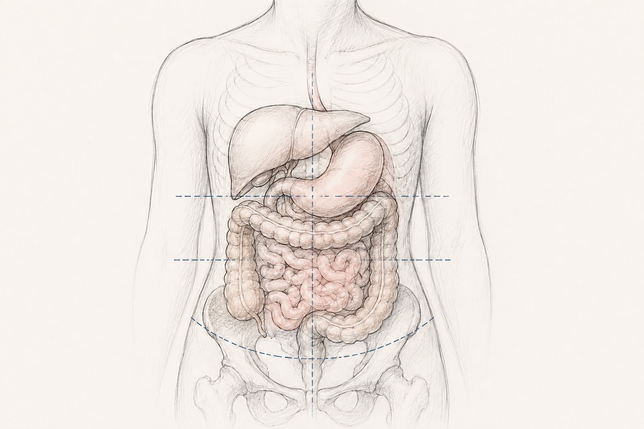
“Viscera” refers to the internal organs of the abdominal, thoracic, and pelvic cavities. “Ptosis” simply means downward displacement from a structure’s normal anatomical position. Together, visceroptosis describes one or more organs that have descended lower than they anatomically should.
Visceroptosis by Organ System
Historically, different forms of visceroptosis were named according to the affected organ:
- Gastroptosis: downward displacement of the stomach, sometimes extending as far as the pelvic floor.
- Enteroptosis: descent of the small intestine.
- Coloptosis: drooping of the colon, particularly the transverse colon, which has been associated with slow-transit constipation.
- Nephroptosis: descent of the kidney from its normal retroperitoneal position.
- Rectal prolapse: protrusion of the entire rectum through the anus; already recognized as a feature of EDS and included in the 2017 hEDS diagnostic criteria.
- Pelvic organ prolapse (POP): descent of the bladder, uterus, or rectum into or through the vaginal wall. Up to 50% of women in the general population are thought to have some degree of prolapse on examination at some point in their lives. POP appears to be more common and often develops at younger ages in people with EDS, including those without traditional risk factors such as childbirth. Pelvic organ prolapse is recognized within the 2017 hypermobile EDS diagnostic criteria and includes:
- Rectocele: the rectum bulges into the back wall of the vagina.
- Cystocele: the bladder bulges into the front wall of the vagina.
- Enterocele: a loop of small bowel descends between the rectum and the vagina or uterus.
The Early Years of Visceroptosis
A Forgotten Diagnosis
In 1885, French physician Dr. Glénard described a condition in which his patients’ abdominal organs sat dramatically lower than expected. Other physicians soon reported similar findings, and the condition became known as “Glénard’s disease.” Over time, medicine shifted to the descriptive anatomical term visceroptosis.
For decades, visceroptosis was considered a legitimate subject of medical inquiry. Radiologists looked for it, surgeons attempted to treat it, and it appeared regularly in major medical journals, including The New England Journal of Medicine, JAMA, The BMJ, and The Lancet.
Then, it quietly disappeared, falling out of clinical fashion for reasons the medical literature never fully explains.
More than half a century later, visceroptosis began reappearing in the literature, primarily through case reports in one particular patient population: Ehlers-Danlos syndrome.
This is the story of a condition that was once recognized, then largely forgotten, and is only now beginning to receive renewed attention, while the people living with its consequences never stopped searching for answers.
What Early Physicians Observed
Descriptions published between 1885 and the 1930s painted a remarkably consistent clinical picture. Physicians repeatedly described:
- Predominantly younger, thin female patients.
- A published lecture from 1911 even suggested organ ptosis was up to ten times more common in women than men.
- Fatigue, pain, and gastrointestinal symptoms that often appeared multisystemic in nature.
- Diagnoses such as neurasthenia and hysteria rather than an investigation of any underlying structural cause.
At the time, these patterns were not recognized as potentially stemming from an underlying structural issue. Instead, these “nervous” characteristics were often treated as the primary problem, while the displaced organs were viewed as incidental or even as consequences of psychological distress rather than possible causes of physical symptoms.
Looking back today, the overlap with modern hEDS is difficult to ignore. Patients with hEDS are disproportionately women, commonly experience pain, fatigue, and gastrointestinal symptoms, and frequently report having those symptoms attributed to anxiety before structural explanations are explored. While the terminology has changed from “neurasthenic” to “anxious” or “somatic,” the pattern of dismissal remains strikingly familiar.
Visceroptosis Today
Anatomic vs. Clinical Visceroptosis: Why the Distinction Matters
The most important distinction is the difference between anatomic and clinical visceroptosis. An organ may sit lower than expected without causing symptoms. In clinical visceroptosis, however, that same displacement interferes with normal physiology and produces meaningful symptoms. The degree of organ descent alone does not determine whether someone is affected. This principle was recognized as early as 1930 in a review published in Radiology, which emphasized that a diagnosis of clinical visceroptosis is appropriate only after other organic disease has been excluded and the patient’s symptoms can reasonably be attributed to the displaced organ rather than simply to the extent of its descent.
Symptoms
Symptoms vary depending on which organs are affected. Pelvic organ prolapse and rectal prolapse commonly cause pelvic pressure, a vaginal or rectal bulge, urinary symptoms, fecal incontinence, or difficulty with bowel emptying. By contrast, the most frequently reported symptoms in symptomatic abdominal visceroptosis are severe constipation and bloating, along with abdominal pain, early satiety, nausea, and gastroparesis-like symptoms.
What Modern Literature Shows
Evidence of visceroptosis in EDS is still very limited. However, some forms of visceroptosis are already well recognized in hEDS. For example, rectal prolapse and pelvic organ prolapse are included among the 2017 diagnostic criteria for hEDS. A few small case reports and studies have also described gastroptosis, transverse colon ptosis, and multiple visceral ptoses in some patients with hEDS. A larger 2023 study of 139 patients with slow-transit constipation and 125 healthy controls found significantly greater transverse colon ptosis in the constipation group, with more severe ptosis associated with a greater likelihood of requiring surgery. Although this study was not conducted in an EDS population, it demonstrates that organ descent may be clinically significant and associated with measurable disease severity.
While these studies are encouraging, an evidence-based relationship between connective tissue laxity, the degree of organ prolapse, and symptom severity remains uncertain. For example, gastrointestinal problems in hEDS are considered multifactorial and may involve disorders of gut–brain interaction, altered motility, autonomic dysfunction, pelvic floor dysfunction, and other structural abnormalities. Because symptoms such as constipation, bloating, abdominal pain, nausea, and early satiety can arise through several of these mechanisms, it is often difficult to determine whether an abnormally positioned organ is causing symptoms, contributing to them, or simply represents an incidental anatomical finding.
Why Diagnosis Remains Challenging
While conditions such as rectal prolapse, pelvic organ prolapse, and nephroptosis have established diagnostic pathways, the same is not true for many other forms of visceroptosis. Standardized diagnostic criteria and imaging protocols are lacking, so these conditions are often not considered or investigated. Standard gastrointestinal evaluations are designed to identify motility disorders or diseases of the digestive tract, not whether abdominal organs have shifted from their normal position.
Where Treatment Stands Today
Conservative management remains the first line of treatment for many forms of visceroptosis. Depending on the individual and type of visceroptosis, this may include dietary modifications, medications, pelvic floor physical therapy, strengthening of the abdominal and back muscles, or abdominal compression garments. For example, pelvic organ prolapse is often managed initially with pelvic floor physical therapy, stabilizing and strengthening the muscles that support the pelvic organs.
When conservative management fails, surgery may be considered. However, while conditions such as rectal prolapse, nephroptosis, and pelvic organ prolapse have established treatment pathways, surgery for other forms of symptomatic abdominal visceroptosis in people with EDS remains poorly studied. The available evidence is extremely limited, and while anecdotally some patients report improvement, others experience little benefit or worsening symptoms. In people with EDS, connective tissue fragility may also complicate surgical planning and long-term outcomes, making individualized treatment decisions particularly important. Any decisions regarding surgery should be made carefully, taking into account symptom severity and the available evidence.
Key Takeaways
- Visceroptosis is a term used to describe the downward movement of one or more internal organs from their normal position.
- Once widely recognized, visceroptosis gradually disappeared from medical practice despite never being definitively disproven.
- Different forms of visceroptosis include gastroptosis, enteroptosis, coloptosis, nephroptosis, rectal prolapse, and pelvic organ prolapse.
- Anatomic and clinical visceroptosis are not the same thing. Not all organ prolapses are clinically significant. Some people may naturally have organs that sit lower than expected without causing symptoms.
- Some forms of visceroptosis, such as rectal prolapse and pelvic organ prolapse, have established diagnostic and treatment pathways.
- Rectal and pelvic organ prolapse are recognized features of hEDS and included in the 2017 hEDS diagnostic criteria.
- By contrast, other forms of visceroptosis in EDS remain poorly understood and poorly researched, with currently no standardized diagnostic criteria, imaging guidelines, or treatment pathways. Therefore, whether connective tissue laxity in EDS contributes to prolapse of abdominal organs, and if these prolapses are clinically significant enough to cause symptoms, remains an active area of research. Greater awareness and larger, high-quality studies are needed.
Edited by Jacqueline Teti, Editor-in-Chief &
Director of Programs, CPP
July, 2026