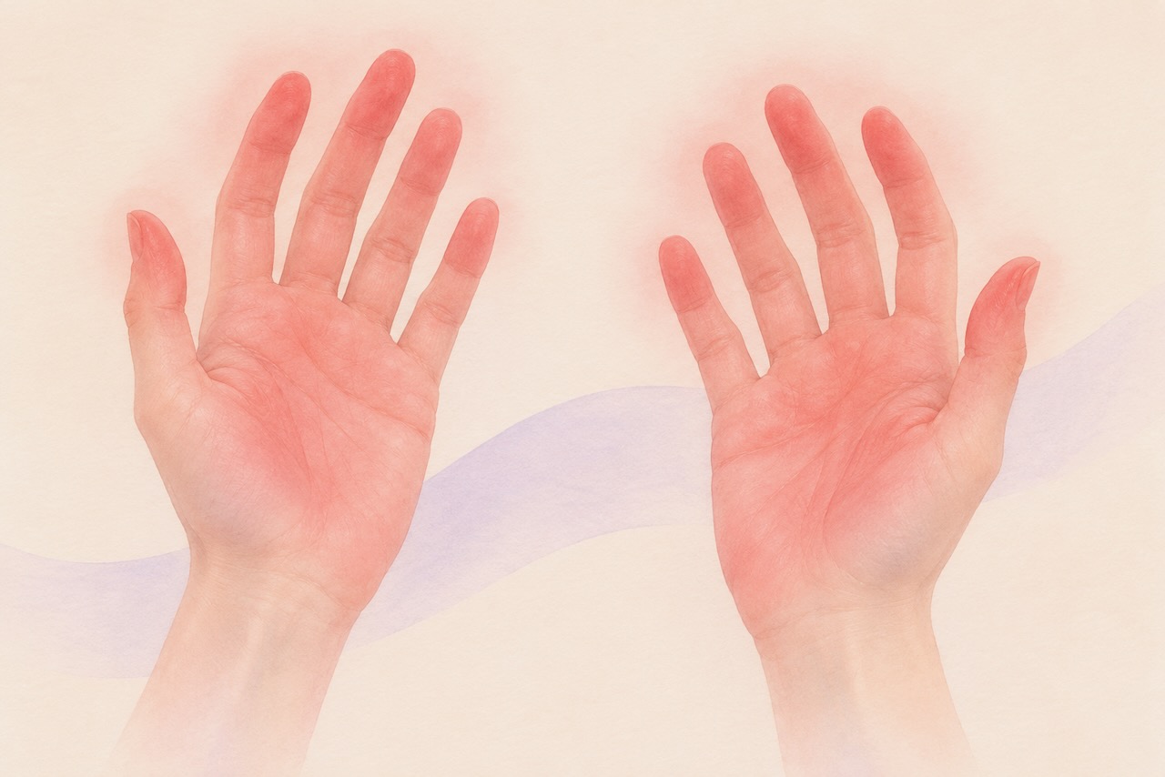
For people living with hypermobile Ehlers-Danlos syndrome (hEDS) and related hypermobility disorders (like HSD), noticing redness in the hands or feet may not be unusual. Dysautonomia, a dysfunction of the autonomic nervous system that regulates automatic body processes like heart rate, blood pressure, and circulation, can be common in hEDS and HSD. One of its most visible manifestations is blood pooling, which can cause the hands and feet to take on a pink or purplish color when lowered.
But not all redness in the extremities is blood pooling. A rare condition called erythromelalgia produces redness that looks superficially similar, but behaves very differently, and for some people with hEDS, understanding that difference matters.
Erythromelalgia is increasingly understood to be a dysautonomia syndrome in its own right, sharing biological mechanisms with conditions already familiar to the hEDS community. Some recent research has found that the pattern of autonomic nerve fiber loss seen in erythromelalgia closely mirrors what is observed in both small fiber neuropathy (SFN) and postural orthostatic tachycardia syndrome (POTS), two conditions with well-established connections to hEDS and HSD. In secondary erythromelalgia specifically, this autonomic nerve fiber loss impairs blood flow regulation, leading to excessive hyperemia (increased blood flow and redness) in response to heat. For people navigating the overlapping landscape of dysautonomia, SFN, and hEDS, recognizing that erythromelalgia may be part of that same biological family is an important step toward understanding what may be happening in their own bodies.
What is Erythromelalgia?
Erythromelalgia is a rare condition characterized by a triad of redness, warmth, and burning pain, ranging from mild to severe. It most commonly affects the feet, occurring in approximately 90% of patients, with the hands and fingers involved in around 25% of cases. In rare circumstances it can affect the face, neck, or other areas of the body. Although it typically presents bilaterally, it can occur unilaterally. Episodes can often begin with itching and mild tingling and progress to an intense burning sensation, which can last minutes to days. These are typically triggered by warmth, exercise, standing, stress, alcohol, caffeine, or spicy food, and are often worse at night. Between episodes, the affected area typically returns to normal with no redness, warmth and burning pain.
Types of Erythromelalgia
Erythromelalgia is classified into two main categories: primary and secondary.
Primary erythromelalgia, the most common type, can be idiopathic, meaning it occurs for unknown reasons, or inherited. The inherited form is caused by gain-of-function mutations (which can either introduce a new property or hyperactivate a biological process) in genes encoding voltage-gated sodium channels, specifically the SCN9A, SCN10A, and SCN11A genes. This makes pain-sensing nerve fibers hyperexcitable, causing them to fire in response to stimuli that would not normally be painful.
Secondary erythromelalgia occurs in association with an underlying condition, such as myeloproliferative disorders (blood conditions involving abnormal cell production). There is also increasing evidence that erythromelalgia may be related to peripheral neuropathies (such as SFN). The most common causes of neuropathies include:
- autoimmune diseases, such as Sjögren’s disease and lupus
- diabetes
- certain medications, including some chemotherapy drugs
- Vitamin B12 deficiency (malabsorption is more common in hEDS and HSD due to numerous comorbid gastrointestinal conditions)
In secondary erythromelalgia, the symptoms of patients tend to be milder than in primary erythromelalgia, and treating the underlying condition may improve or resolve the symptoms.
Erythromelalgia vs. Blood Pooling: How to Tell the Difference
This distinction is particularly important for the hEDS community, where blood pooling from dysautonomia is common and can be mistaken for erythromelalgia, or vice versa.
Blood pooling can occur when the autonomic nervous system fails to properly regulate circulation, allowing blood to accumulate in the hands or feet when they are lowered. This produces a pink, red, or purplish discoloration, sometimes called acrocyanosis when it takes on a blue-purple hue, that is typically painless or mildly uncomfortable. Crucially, the discoloration of blood pooling improves when the affected area is elevated. Lying down, for example, typically restores normal color to the feet relatively quickly. For the hands, raising them above heart level can have a similar effect.
Erythromelalgia behaves differently in several important ways. While lifting the affected limb can ease the intensity of symptoms during a flare, the redness does not reliably resolve with elevation alone. It is typically accompanied by warmth and burning discomfort or pain, rather than discoloration alone.
Erythromelalgia is also triggered or worsened by heat, while blood pooling is primarily positional (although it can be more pronounced in hot environments).
These conditions are not mutually exclusive, and many individuals can have both. A practical way to think about the distinction is that if the redness in your hands and feet goes away when elevated and there is no accompanying burning, it is more likely blood pooling than erythromelalgia.
The Connection to Small Fiber Neuropathy and EDS
The link between erythromelalgia and SFN, a condition in which the small nerve fibers responsible for pain, temperature, and autonomic regulation are damaged or lost, is increasingly well supported by research. SFN is now recognized as a significant and common feature of hEDS and HSD, with studies finding evidence of small fiber involvement in the majority of patients evaluated (Chronic Pain Partners covered this topic here).
A small 2025 study examining the skin biopsy findings of patients with erythromelalgia found they had damaged and weakened nerves of their sweat glands and arteries of the affected tissues, which mirrors the effect seen in SFN. The study proposed that erythromelalgia may represent a dysautonomia syndrome, characterized by vascular changes and nerve fiber loss that overlap significantly with what is seen in SFN. This finding adds biological plausibility to the idea that erythromelalgia and SFN share underlying mechanisms, and that people with hEDS and HSD, who already have a higher prevalence of SFN and dysautonomia, may be at greater risk for erythromelalgia than the general population.
Diagnosis
There is no single definitive test for erythromelalgia. Diagnosis is based on clinical history, physical examination during an active episode or photographs of flares, and ruling out other conditions. Because episodes come and go, only about two-thirds of patients have visible findings at the time of a clinical appointment, making a detailed symptom history essential.
Investigations may include blood tests to rule out myeloproliferative disorders, genetic testing for known sodium channel mutations in suspected inherited cases, and thermography, a specialized imaging technique that maps differences in skin temperature. Electromyography (EMG) and nerve conduction studies may be used to assess for other nerve-related disorders, but they are usually normal in isolated SFN and cannot rule it out. The quantitative sudomotor axon reflex test (QSART) can also help evaluate underlying nerve dysfunction in people with erythromelalgia by assessing the function of small autonomic nerve fibers that control sweating. Although it can provide supporting evidence of autonomic small-fiber dysfunction, it cannot diagnose erythromelalgia on its own.
For people with hEDS who are experiencing episodes of intense burning redness in the extremities, raising the possibility of erythromelalgia with a neurologist or dermatologist is a well-supported step. The condition is rare and can be missed, particularly in patients whose primary diagnosis is already associated with redness due to dysautonomia.
Management
Erythromelalgia can be challenging to treat, and management requires an individualized approach. The primary goal is identifying and avoiding triggers, the most common of which may include warmth, exercise, alcohol, caffeine, and spicy foods. Non-medical strategies include using a fan, applying cool gel packs wrapped in a thin towel, elevating the affected area, and resting on a cool surface. However, while cooling brings relief, immersing the affected area in ice water should be avoided. Ice baths can cause serious skin damage, including ulceration and necrosis, that is difficult to treat.
For secondary erythromelalgia, treating the underlying condition, including, where applicable, managing SFN, may improve or eliminate symptoms. For example, if vitamin B12 deficiency is an underlying cause, correcting the deficiency may help relieve symptoms.
Medications used for primary and secondary erythromelalgia include anticonvulsants such as gabapentin and carbamazepine, antidepressants such as amitriptyline and venlafaxine, aspirin for cases associated with blood disorders, and topical treatments including lidocaine patches and capsaicin cream. None of these treatments are fully curative, but many patients find meaningful relief with the right combination.
Key Takeaways
- Erythromelalgia is a rare condition characterized by a triad of redness, warmth, and burning pain, ranging from mild to severe, that most commonly affects the hands and feet.
- It is distinct from blood pooling associated with dysautonomia, where blood pooling typically improves with elevation and is not accompanied by burning pain, while erythromelalgia does not reliably resolve with elevation and is relieved by cooling rather than position change.
- Erythromelalgia seems to be linked to small fiber neuropathy SFN), which is itself a recognized and common feature of hEDS and HSD.
- Some recent research suggests erythromelalgia may represent a dysautonomia syndrome involving autonomic nerve fiber loss that mirrors what is seen in SFN.
- Diagnosis is clinical, based on symptom history and episode characteristics, and may require blood tests, genetic testing, QSART, or thermography to confirm and rule out secondary causes.
- Management focuses on trigger avoidance, cooling strategies, and medications. Ice baths should be avoided as they can cause serious skin damage.
- For secondary erythromelalgia associated with SFN, treating an underlying cause of the neuropathy, such as vitamin B12 deficiency, may improve or resolve symptoms.
- People with hEDS experiencing unexplained episodes of intense burning redness in the extremities should consider raising the possibility of erythromelalgia with a neurologist or dermatologist.
Final Thoughts
For people living with hEDS and HSD, the body’s signals can be difficult to interpret, particularly when several conditions overlap and share similar features. Erythromelalgia is one condition that may be hiding in plain sight, mistaken for the blood pooling that many in this community already know and manage.
Understanding the difference between these two conditions is important as it opens the door to the right diagnosis, and the right management strategies. While erythromelalgia presents real challenges, many people live full and active lives with this condition, and the growing recognition of its connection to SFN and dysautonomia is bringing it closer to the center of clinical attention it deserves.
By Jacqueline Moltzau Anderson, PhD
Researcher at University of California
November, 2026
If you like what you read, please comment and share!



Hello. I have very severe erythromelalgia. It would last from October until May. My skin on my hands and feet would actually blister under the skin. I would lose toenails and fingernails from it. I was referred to a pain doctor and she prescribed low-dose naltrexone for it. I started slow at 0.5 mg and am now up to 5 mg. It has been almost a year and it has not come back. It was so painful I was ready to give up on life. I thank God for that doctor and LDN. It gave me my life back.
I have H-EDS, POTS, MCAS and dysautonomia ++++