Exciting news from Poland! Researchers from the Department of Clinical Genetics at Nicolaus Copernicus University in Torún, Poland, released a preprint of research that may hold the answers to the cause of hypermobile Ehlers-Danlos Syndrome (hEDS) for a part of the patient population. Specifically, Junkiert-Czarnecka et al. investigated the MIA3 gene in people with hEDS to see if variants or mutations of this gene could be the cause or a part of the mechanism for some of the hEDS cases.
The researchers included 100 patients with clinically-diagnosed hEDS who also tested negative for other connective tissue disorders, like other forms of EDS, Marfan syndrome, and more. They analyzed all of these patients for changes in the MIA3 gene which encodes a protein that helps to create the extracellular matrix.
At the molecular level
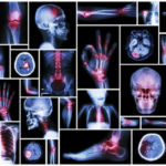
Between the cells that make up the tissues in the human body, two different structures exist: the interstitial fluid and the extracellular matrix (ECM). The interstitial fluid is an area between the cells that contains nutrients, water, proteins, hormones, wastes, and more. Its purpose is to deliver nutrients from the blood vessels to the cells. The extracellular matrix shares this space and is made up of elastin fibers, collagen fibers, glycoproteins, and proteoglycans. Depending on the tissue type, the interstitial fluid and the ECM can have different functions. In some tissues, the ECM connects to the inside of the cells, holding the tissue together while creating structure and support. Since the ECM contains collagen, this is where the connection to hEDS may come into play.
The collagen that makes up the ECM is created inside the cells. There are two delivery systems for collagen molecules. The one this research focuses on is the vesicles–they help take the collagen molecules from inside the cells to the ECM. The vesicles are also made up of a collagen-like material. The gene MIA3 encodes the protein TANGO1. TANGO1 provides instructions that assist in making the vesicles. If TANGO1 is shortened, the instructions for making the vesicles aren’t available. If the vesicles aren’t available, collagen and other proteins can’t be delivered to the ECM. Therefore, the ECM isn’t as strong or supportive due to the vesicles not being able to deliver the collagen created inside of the cells. In fact, in previous research, patients with joint hypermobility syndrome and hEDS showed that many items researchers expected to see in the ECM, including collagen, were inside the cell’s cytoplasm instead. The reason for this was not explored in this previous research, but MIA3 and TANGO1 may hold the answer.
Results
Researchers found structural changes in the MIA3 gene in 14 of the 100 recruited hEDS patients. Thirteen of those changes were likely benign, but one of the variants truncated the TANGO1 protein. This was in a 49-year-old woman who scored a seven out of nine on the Beighton scale and has a number of other symptoms associated with hEDS. This woman has a 14-year-old daughter who then had her genetics tested. The daughter, who has similar symptoms of hEDS, with a score of nine out of nine on the Beighton scale, also showed the same variant in the MIA3 gene and the truncation of the TANGO1 protein.
Without the TANGO1 protein, this participant and her daughter would not have been able to make the vesicles necessary to transport collagen to the ECM. The researchers said that since only one participant out of 100 hundred in their study showed this change–and all participants had clinical signs of hEDS–more research needs to be done, as the truncated TANGO protein on altered MIA3 genes may be just part of one potential mechanism of hEDS.
Previous hEDS genetic research
Other possibilities for the genetic cause of hEDS have been identified and published in the recent past.
In 2015, several members across generations in a Belgian family showed a variant of LZST1. This was found in the family members with clinical diagnoses of hEDS but not in any of the unaffected family members. However, variants in this gene are usually associated with cancer, not connective tissue creation or transport. Further research is needed to find out if this gene and its variants play a role in causing hEDS.
In 2016, researchers found that alterations in TPSAB1 increased basal serum tryptase levels in 35 families. These families had, among other symptoms, dysautonomia, GI disorders, and connective tissue abnormalities. However, not all affected individuals met diagnostic criteria, and elevated levels of basal serum tryptase are relatively common in the general population.
The TNXB gene is next on the list of possible causes for hEDS. TNXB encodes Tenascin X, which is a protein important for collagen organization in the extracellular matrix. Tenascin X deficiency has been shown to cause a recessive version of EDS, currently classified as clEDS. TNXB haploinsufficiency (where one copy of a gene is deleted or inactivated, and the remaining gene cannot provide or produce enough for normal functioning) has been present in a small subset of patients with hEDS.
In other cases, patients with congenital adrenal hyperplasia (CAH) may have a mutation of the CYP21A2 gene and the neighboring TNXB. This can lead to both CAH and hEDS, a unique EDS type which is called CAH-X. More research will need to be done to see if and how TNXB and other genes work individually or together to cause hEDS.
EDS Awareness has previously interviewed Dr. Russell “Chip” Norris of the Norris Lab at the Medical University of South Carolina, a lab dedicated to research on connective tissue diseases and abnormalities, including EDS. In 2018, Dr. Courtney Genesmer, then a first-year research student, was interested in studying hEDS due to her own experiences as a patient with hEDS. Her involvement transformed the lab into “one of the world’s largest groups studying hEDS.”
The Norris Lab researchers have produced a review of the scientific literature on hEDS and continue to research the genetic cause of hEDS. They recently identified a “strong candidate” for the cause of hEDS (results haven’t been published but are forthcoming) and continue to research as they believe that “hEDS likely involves multiple genes.”
In addition, the Ehlers-Danlos Society has enrolled a thousand people in its HEDGE study to expedite finding the cause of hEDS.
Conclusion
Finding the genetic mechanism behind hypermobile Ehlers-Danlos Syndrome would provide answers to a long-standing question in the EDS communities. It could lead to more specific diagnostic criteria, up to and including genetic testing. With genetic testing, patients with clinical diagnoses would be less likely to be questioned about whether their diagnosis is accurate. A specific mutation on a genetic test removes the ambiguity many hEDS patients currently face. Additionally, multiple genetic sources of hEDS may also lead to the diagnosis of “hypermobile EDS” being broken into more specific subtypes. Most importantly of all, knowing the reason for hEDS could lead to more treatments and therapies for patients with the condition. The world continues to inch closer to an answer to the biggest mystery plaguing the EDS community.
Cover Image by National Institute of Cancer (Unsplash)
Kate Schultz
April 2023


I wish someone would use my family’s DNA. I scored an 8/9 at age 45. My (at the time) 5 year old son scored a 9/9, and my 21 y/o son scored a 9/9. I have a, now 9 y/o daughter who we are almost positive has heads, waiting on Genetics to make it official. That’s 3/3 children.
Wow, it does sound like EDS runs in your family. You may want to reach out to the Norris lab and see if your family’s DNA would be helpful in their studies: https://www.thenorrislab.com/
I have the MIA3 mutation & I have hEDS. I was initially dx’d with Marfan’s at age 20 before they had genetic testing & then told yrs later that I did not have Marfan’s & that was it. I was correctly dx’d in my late 40’s w/hEDS after severe medical issues & am now disabled & no longer able to work. My grandfather died of an aortic aneurysm & I have one but am being monitored by cardiology. I think it’s important to get more people tested w/hEDS & figure out what genes are involved so people can be properly diagnosed. There is so much we still do not know.